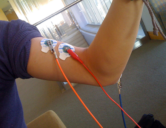
Browse Articles

The use of computer vision to differentiate valley fever from lung cancer via CT scans of nodules

El Kereamy et al. | Nov 12, 2024

The use of computer vision to differentiate valley fever from lung cancer via CT scans of nodules

Pulmonary diseases like lung cancer and valley fever pose serious health challenges, making accurate and rapid diagnostics essential. This study developed a MATLAB-based software tool that uses computer vision techniques to differentiate between these diseases by analyzing features of lung nodules in CT scans, achieving higher precision than traditional methods.

Read More...

Enhancing marine debris identification with convolutional neural networks

Wahlig et al. | Apr 03, 2024

Enhancing marine debris identification with convolutional neural networks
Image credit: The authors

Plastic pollution in the ocean is a major global concern. Remotely Operated Vehicles (ROVs) have promise for removing debris from the ocean, but more research is needed to achieve full effectiveness of the ROV technology. Wahlig and Gonzales tackle this issue by developing a deep learning model to distinguish trash from the environment in ROV images.

Read More...

The Role of a Mask - Understanding the Performance of Deep Neural Networks to Detect, Segment, and Extract Cellular Nuclei from Microscopy Images

Dasgupta et al. | Jul 06, 2021

The Role of a Mask - Understanding the Performance of Deep Neural Networks to Detect, Segment, and Extract Cellular Nuclei from Microscopy Images

Cell segmentation is the task of identifying cell nuclei instances in fluorescence microscopy images. The goal of this paper is to benchmark the performance of representative deep learning techniques for cell nuclei segmentation using standard datasets and common evaluation criteria. This research establishes an important baseline for cell nuclei segmentation, enabling researchers to continually refine and deploy neural models for real-world clinical applications.

Read More...

Identification of microwave-related changes in tissue using an ultrasound scan

Shariff et al. | Apr 24, 2024

Identification of microwave-related changes in tissue using an ultrasound scan
Image credit: Shariff and Shariff 2024

Microwave energy (ME) is used in the medical field to denature protein structures, resulting in inactivation or destruction of abnormal cells. Identifying the extent of destruction of abnormal tissue (cancer tissue or tissue with abnormal electrical activity) is essential for accomplishing successful therapy and reducing collateral damage. Our study was an ex vivo assessment of the changes on ultrasound scans (US) in chicken tissue exposed to ME. We hypothesized that any changes in tissue structures would be recognized on the reflected ultrasound waves. Ultrasound scans of tissues change with exposure to microwaves with increasing reflection of ultrasound waves. With exposure to microwaves, surface level brightness on the ultrasound scans increases statistically significantly. The findings could be used in heat related (ME and radiofrequency) procedures where clinicians would be able to actively assess lesions in real-time. Further studies are required to assess changes in tissue during active exposure to different types of energies.

Read More...

The Role of Temporal Lobe Epilepsy in Cardiac Structure and Function

Choi et al. | Aug 15, 2018

The Role of Temporal Lobe Epilepsy in Cardiac Structure and Function

Cardiac autonomic and structural changes may occur in temporal lobe epilepsy patients and contribute to the risk of sudden unexpected death in epilepsy patients. Choi and colleagues reviewed clinical charts to obtain patients’ lifetime seizure count, antiepileptic drug use, and history of heart disease, followed by transthoracic echocardiogram to calculate left ventricle dimensions, ejection fraction, and left ventricle mass. By comparing epilepsy patients to control subjects, they found that epilepsy patients had thinner left ventricle walls and smaller ejection fraction, but with no significant difference in left ventricle mass.

Read More...