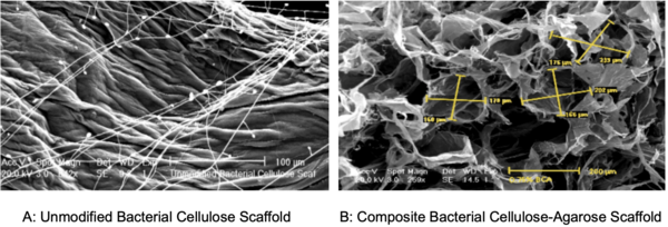
In this study, the authors investigate the suitability of using bacterial cellulose as a scaffold for cell transplants. Interestingly, this cellulose is a can be found in the discard from a symbiotic culture of bacteria and yeast (SCOBY) used to make kombucha.
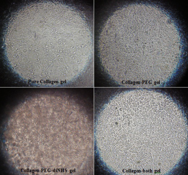
Environment affects the progression of life, especially at the cellular level. This study investigates multiple 3-dimensional growth environments, also known as scaffolds or hydrogels, and their effect on the growth of a type of cells called fibroblasts. These results suggest that a scaffold made of collagen and polyethylene glycol are favorable for cell growth. This research is useful for developing implantable devices to aid wound healing.
In this study, the authors develop a new hydrogel using photochemical crosslinking with bovine serum albumin and methylene blue. They find that this new hydrogel has some useful applications!
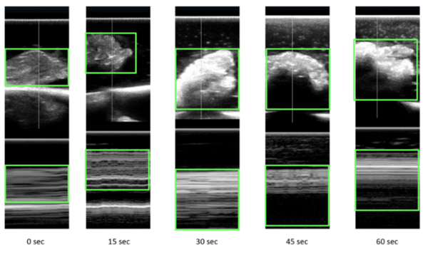
Microwave energy (ME) is used in the medical field to denature protein structures, resulting in inactivation or destruction of abnormal cells. Identifying the extent of destruction of abnormal tissue (cancer tissue or tissue with abnormal electrical activity) is essential for accomplishing successful therapy and reducing collateral damage. Our study was an ex vivo assessment of the changes on ultrasound scans (US) in chicken tissue exposed to ME. We hypothesized that any changes in tissue structures would be recognized on the reflected ultrasound waves. Ultrasound scans of tissues change with exposure to microwaves with increasing reflection of ultrasound waves. With exposure to microwaves, surface level brightness on the ultrasound scans increases statistically significantly. The findings could be used in heat related (ME and radiofrequency) procedures where clinicians would be able to actively assess lesions in real-time. Further studies are required to assess changes in tissue during active exposure to different types of energies.
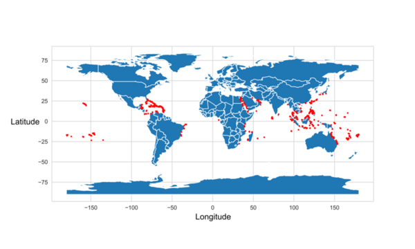
Coral bleaching is a fatal process that reduces coral diversity, leads to habitat loss for marine organisms, and is a symptom of climate change. This process occurs when corals expel their symbiotic dinoflagellates, algae that photosynthesize within coral tissue providing corals with glucose. Restoration efforts have attempted to repair damaged reefs; however, there are over 360,000 square miles of coral reefs worldwide, making it challenging to target conservation efforts. Thus, predicting the likelihood of bleaching in a certain region would make it easier to allocate resources for conservation efforts. We developed a machine learning model to predict global locations at risk for coral bleaching. Data obtained from the Biological and Chemical Oceanography Data Management Office consisted of various coral bleaching events and the parameters under which the bleaching occurred. Sea surface temperature, sea surface temperature anomalies, longitude, latitude, and coral depth below the surface were the features found to be most correlated to coral bleaching. Thirty-nine machine learning models were tested to determine which one most accurately used the parameters of interest to predict the percentage of corals that would be bleached. A random forest regressor model with an R-squared value of 0.25 and a root mean squared error value of 7.91 was determined to be the best model for predicting coral bleaching. In the end, the random model had a 96% accuracy in predicting the percentage of corals that would be bleached. This prediction system can make it easier for researchers and conservationists to identify coral bleaching hotspots and properly allocate resources to prevent or mitigate bleaching events.
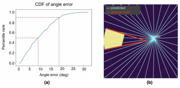
Robot-assisted minimally invasive surgery (RMIS) benefits from increased precision and faster recovery, with force feedback from the surgical tool being critical for control. Researchers tested the use of neural networks for detecting the vanishing point of the tool, a key element for force feedback.
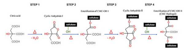
Hydrogels are commonly used in medicine, pharmaceuticals, and agriculture. Hydrogels absorb water by swelling and re-release this water by diffusion. This study sought to synthesize a biodegradable, cellulose-based hydrogel that is more effective at absorbing and re-releasing water than those produced by current methods. We tested the compressive strength of both the dry and swollen gels and the tensile strength of the swollen gels to elucidate the gel structure.