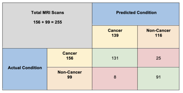
AI analysis of brain scans offers promise for helping doctors diagnose brain tumors. Haider and Drosis explore this field by developing machine learning models that classify brain scans as "cancer" or "non-cancer" diagnoses.
Read More...The utilization of Artificial Intelligence in enabling the early detection of brain tumors
AI analysis of brain scans offers promise for helping doctors diagnose brain tumors. Haider and Drosis explore this field by developing machine learning models that classify brain scans as "cancer" or "non-cancer" diagnoses.
Read More...Unlocking robotic potential through modern organ segmentation
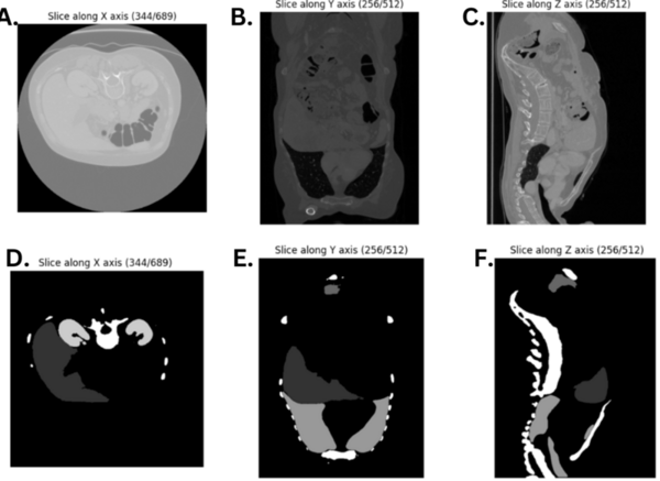
The authors looked at different models of semantic segmentation to determine which may be best used in the future for segmentation of CT scans to help diagnose certain conditions.
Read More...Functional Network Connectivity: Possible Biomarker for Autism Spectrum Disorders (ASD)
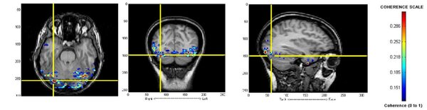
Autism spectrum disorder (ASD) is a complex neurodevelopmental disorder and is difficult to diagnose in young children. Here magnetoencephalography was used to compare the brain activity in patients with ASD to patients in a control group. The results show that patients with ASD have a high level of activity in different areas of the brain than those in the control group.
Read More...Large-scale brain network connectivity under anxiety induced by naturalistic story listening
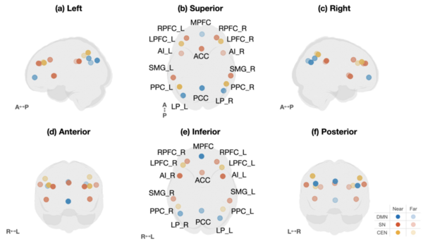
This study found that anxiety induced by a suspenseful story increased communication between the brain’s salience, default mode, and central executive networks, with the central executive network acting as a bridge during peak tension. These findings suggest that anxiety alters large-scale brain connectivity patterns and may help inform future diagnostic tools and personalized treatments for anxiety disorders.
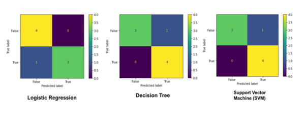
Read More...Assessing machine learning model efficacy for brain tumor MRI classification: a multi-model approach
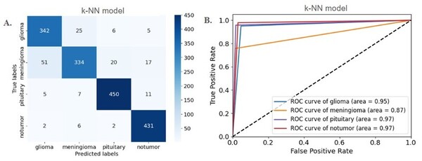
This manuscript explores the performance of five different machine learning models in classifying brain tumors from a dataset of MRI scans. The authors find that several of the models showed >90% accuracy. Thus, the authors suggest that machine learning models demonstrate potential for effective implementation in clinical settings, including as a diagnostic tool that can be used to complement the expertise of neuroradiologists.
Read More...Towards multimodal longitudinal analysis for predicting cognitive decline

Understanding and predicting cognitive decline in Alzheimer's disease
Read More...Using satellite surface temperature data to monitor urban heat island
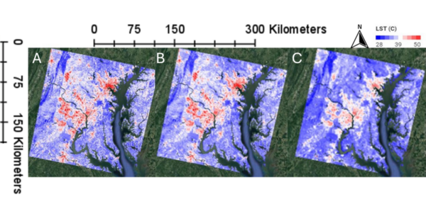
This manuscript investigates the urban heat island (UHI) effect by utilizing two satellite datasets: Landsat (high spatial resolution, lower temporal resolution) and MODIS (lower spatial resolution, high temporal resolution). The authors hypothesized that Landsat would provide better spatial detail, while MODIS would better capture temporal variations. Their analysis in the Washington D.C.–Baltimore region supports these hypotheses, demonstrating that Landsat offers finer spatial details, whereas MODIS provides more consistent seasonal patterns and better detects heatwave frequencies.
Read More...Studying the effects of different anesthetics on quasi-periodic patterns in rat fMRI
The authors looked at the effects of commonly used anesthetics in rodents on brain activity (specifically quasi-periodic patterns). Understanding effects on brain activity is important for researchers to understand when choosing rodent models for disease.
Read More...Transfer Learning with Convolutional Neural Network-Based Models for Skin Cancer Classification
Skin cancer is a common and potentially deadly form of cancer. This study’s purpose was to develop an automated approach for early detection for skin cancer. We hypothesized that convolutional neural network-based models using transfer learning could accurately differentiate between benign and malignant moles using natural images of human skin.
Read More...A machine learning approach to detect renal calculi by studying the physical characteristics of urine
The authors trained a machine learning model to detect kidney stones based on characteristics of urine. This method would allow for detection of kidney stones prior to the onset of noticeable symptoms by the patient.
Read More...