Assessing machine learning model efficacy for brain tumor MRI classification: a multi-model approach
(1) Lexington High School
https://doi.org/10.59720/25-127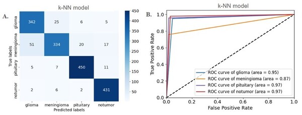
Brain tumors pose a significant diagnostic challenge due to their complexity and the shortage of specialized neuroradiologists, motivating the development of machine learning (ML) approaches to reduce diagnostic variability and augment clinical workflows. Here, we evaluated the effectiveness of ML models in classifying brain tumor types, addressing variability and time-intensive manual interpretation. We investigated two hypotheses. First, we hypothesized that the classification accuracy of ML models would vary across tumor types due to differences in their imaging interpretability. Second, we hypothesized that tumor boundaries and high-intensity regions in MRI scans would disproportionately influence model predictions because these regions have the most discriminative information for separating tumor classes. We used the publicly available “Brain Tumor MRI Dataset” from Kaggle, consisting of 7,023 MRI slices categorized into glioma, meningioma, pituitary, and non-tumorous cases. We evaluated five ML models: Decision Tree, k-Nearest Neighbors (k-NN), DenseNet, ResNet50, and a custom Convolutional Neural Network (CNN). DenseNet, k-NN, and ResNet50 achieved high accuracies of 0.9361, 0.9125, and 0.9112, respectively. Pituitary and non-tumorous cases reached AUC scores of 1.00, while glioma and meningioma had slightly lower AUC scores of 0.99 and 0.98, respectively. Our analyses identified tumor boundaries and high-intensity MRI regions as key features driving classification, and statistical tests confirmed no significant differences among models (p > 0.05). Our research highlights the potential of ML models to enhance diagnostics by improving accuracy and identifying tumor-specific performance variations, supporting future clinical integration.
This article has been tagged with: