Advancements in glioma segmentation: comparing the U-Net and DeconvNet models
(1) Dublin High School, (2) Radcliffe Department of Medicine, University of Oxford
https://doi.org/10.59720/24-022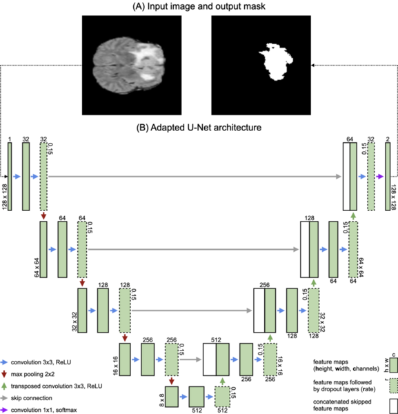
In this study, we address the challenge of accurately segmenting gliomas from magnetic resonance imaging (MRI) scans, an essential task for treatment planning and prognosis in glioma patients. Prompted by the limitations of manual segmentation methods and the need for precise automated techniques, we evaluated the efficacy of advanced deep learning models in this domain. Our primary objective was to compare the performance of two convolutional neural network architectures, the U-Net and a baseline DeconvNet model, in segmenting (outlining) gliomas from surrounding tissues in MRI scans. We hypothesized that the U-Net model would outperform the DeconvNet model in segmenting tumors due to U-Net’s advanced architecture (skip connections). Utilizing the Multimodal Brain Tumor Image Segmentation (BraTS) 2018 dataset for training and validation, we evaluated the models based on the Dice Similarity Coefficient (DSC) to quantify segmentation accuracy. We found that the U-Net model achieved a significantly higher average DSC of 0.918 ± 0.053, compared to 0.867 ± 0.065 for DeconvNet model (p < 0.05), indicating superior accuracy in tumor delineation. Furthermore, the U-Net model showed more stable training and validation losses, suggesting better adaptability to new data. We concluded that the U-Net model’s advanced capabilities enhanced glioma segmentation in MRI scans, surpassing the baseline DeconvNet method. Our findings may help improve non-invasive diagnostic procedures and treatment planning in glioma patients, reinforcing the value of integrating advanced neural network architectures into medical imaging.
This article has been tagged with: