Browse Articles

Effect of hypervitaminosis A in regenerating planaria: A potential model for teratogenicity testing

Bennet et al. | Dec 12, 2022

Effect of hypervitaminosis A in regenerating planaria: A potential model for teratogenicity testing

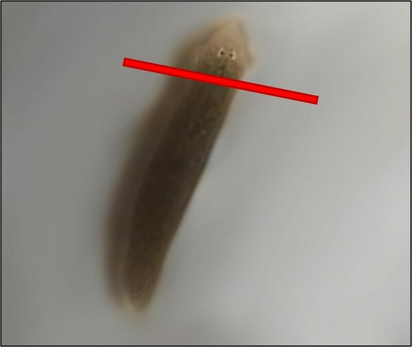
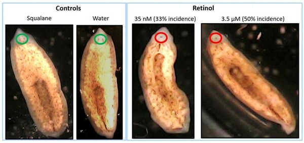
This unique research study evaluated the potential use of the flatworm, brown planaria (Dugesia tigrine), as an alternative model for teratogenicity testing. In this study, we exposed amputated planaria to varying concentrations of a known teratogen, vitamin A (retinol), for approximately 2 weeks, and evaluated multiple parameters including the formation of blastema and eyes. The results from this study demonstrated that high concentrations of retinol caused defects in head and eye formation in regenerating planaria, with similarities to vitamin A related teratogenicity findings in mammals. Based on these results, regenerating brown planaria are a promising alternative model for teratogenicity testing, which can potentially be paradigm shifting as it can reduce cost, time, and pregnant animal use in research.

Read More...
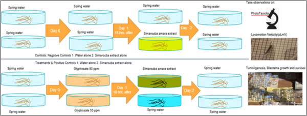
Testing Simarouba amara’s therapeutic effects against weedicide-induced tumor-like morphology in planarians

Thiagarajan et al. | Apr 26, 2024

Testing Simarouba amara’s therapeutic effects against weedicide-induced tumor-like morphology in planarians

According to the World Health Organization, cancer is a leading cause of death globally. The disease’s prevalence is rapidly increasing in association with factors including the increased use of pesticides and herbicides, such as glyphosate, which is one of the most widely used herbicide ingredients. Natural antioxidants and phytochemicals are being tested as anti-cancer agents due to their antiproliferative, antioxidative, and pro-apoptotic properties. Thus, we aimed to investigate the potential role of S. amara extract as a therapeutic agent against glyphosate-induced toxicity and tumor-like morphologies in regenerating and homeostatic planaria (Dugesia dorotocephala).

Read More...

Determining the Effects of Fibroblast Growth Factor 2 on the Regenerative Abilities of Echinometra lucunter Sea Urchins

Kisling et al. | Feb 12, 2019

Determining the Effects of Fibroblast Growth Factor 2 on the Regenerative Abilities of Echinometra lucunter Sea Urchins

As humans, not all our body organs can adequately regenerate after injury, an ability that declines with age. In some species, however, regeneration is a hallmark response that can occur limitless numbers of time throughout the life of an organism. Understanding how such species can regenerate so efficiently is of central importance to regenerative medicine. Sea urchins, unlike humans, can regenerate their spinal tissue after injury. Here the authors study the effect of a growth factor, FGF2, on sea urchin regeneration but find no conclusive evidence for a pro-regenerative effect after spinal tissue injury.

Read More...

The non-nutritive sweeteners acesulfame potassium and neotame slow the regeneration rate of planaria

Russo et al. | Nov 29, 2023

The non-nutritive sweeteners acesulfame potassium and neotame slow the regeneration rate of planaria
Image credit: Russo et al. 2023

The consumption of sugar substitute non-nutritive sweeteners (NNS) has dramatically increased in recent years. Despite being advertised as a healthy alternative, NNS have been linked to adverse effects on the body, such as neurodegenerative diseases (NDs). In NDs, neural stem cell function is impaired, which inhibits neuron regeneration. The purpose of this study was to determine if the NNS acesulfame potassium (Ace-K) and neotame affect planaria neuron regeneration rates. Since human neurons may regenerate, planaria, organisms with extensive regenerative capabilities due to stem cells called neoblasts, were used as the model organism. The heads of planaria exposed to either a control or non-toxic concentrations of NNS were amputated. The posterior regions of the planaria were observed every 24 hours to see the following regeneration stages: (1) wound healing, (2) blastema development, (3) growth, and (4) differentiation. The authors hypothesized that exposure to the NNS would slow planaria regeneration rates. The time it took for the planaria in the Ace-K group and the neotame group to reach the second, third, and fourth regeneration stage was significantly greater than that of the control. The results of this study indicated that exposure to the NNS significantly slowed regeneration rates in planaria. This suggests that the NNS may adversely impact neoblast proliferation rates in planaria, implying that it could impair neural stem cell proliferation in humans, which plays a role in NDs. This study may provide insight into the connection between NNS, human neuron regeneration, and NDs.

Read More...

Wound healing properties of mesenchymal conditioned media: Analysis of PDGF, VEGF and IL-8 concentrations

Prasad et al. | Dec 15, 2021

Wound healing properties of mesenchymal conditioned media: Analysis of PDGF, VEGF and IL-8 concentrations

Regenerative medicine has become a mainstay in recent times, and employing stem cells to treat several degenerative, inflammatory conditions has resulted in very promising outcomes. These forms of cell-based therapies are novel approaches to existing treatment modalities. In this study, the authors compared the concentrations of the cytokines PDGF, IL-8, and VEGF between conditioned and spent media of mesenchymal stem cells (MSCs) to evaluate their potential therapeutic properties for wound healing in inflammatory conditions. They hypothesized that conditioned media contains higher concentrations of wound healing cytokines compared to spent media. The authors found that while IL-8 and VEGF were present in highest concentrations in conditioned media, PDGF was present in maximal amounts in spent media.

Read More...

Specific Transcription Factors Distinguish Umbilical Cord Mesenchymal Stem Cells From Fibroblasts

Park et al. | Aug 16, 2019

Specific Transcription Factors Distinguish Umbilical Cord Mesenchymal Stem Cells From Fibroblasts

Stem cells are at the forefront of research in regenerative medicine and cell therapy. Two essential properties of stem cells are self-renewal and potency, having the ability to specialize into different types of cells. Here, Park and Jeong took advantage of previously identified stem cell transcription factors associated with potency to differentiate umbilical cord mesenchymal stem cells (US-MSCs) from morphologically similar fibroblasts. Western blot analysis of the transcription factors Klf4, Nanog, and Sox2 revealed their expression was unique to US-MSCs providing insight for future methods of differentiating between these cell lines.

Read More...

3D Printed Polymer Scaffolds for Bone Tissue Regeneration

Jayatissa et al. | Apr 26, 2019

3D Printed Polymer Scaffolds for Bone Tissue Regeneration

Scientists are always on the quest to improve the body's healing abilities and broken bones are no exception. In this article, the authors investigate properties of 3D-printed biocompatible polymers used to improve bone healing. With such efforts, we can hope to, one day, improve bone scaffolding materials in ways that make the natural healing processes more efficient, reducing the time needed for recovery from bone fractures.

Read More...

The Effect of Caffeine on the Regeneration of Brown Planaria (Dugesia tigrina)

Lazorik et al. | May 10, 2019

The Effect of Caffeine on the Regeneration of Brown Planaria (<em>Dugesia tigrina</em>)

The degeneration of nerve cells in the brain can lead to pathologies such as Parkinson’s disease. It has been suggested that neurons in humans may regenerate. In this study, the effect of different doses of caffeine on regeneration was explored in the planeria model. Caffeine has been shown to enhance dopamine production, and dopamine is found in high concentrations in regenerating planeria tissues. Higher doses of caffeine accelerated planeria regeneration following decapitation, indicating a potential role for caffeine as a treatment to stimulate regeneration.

Read More...