Utilizing a novel T1rho method to detect spinal degeneration via magnetic resonance imaging
(1) Gilbert Classical Academy, Gilbert, Arizona, (2) Barrow Neurological Institute, Phoenix, Arizona
https://doi.org/10.59720/23-080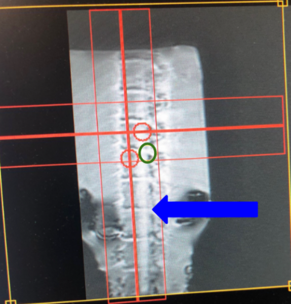
Spinal degeneration has been linked to critical conditions such as osteoarthritis in adults aged 40+; while this condition is considered to be irreversible, we took interest in magnetic resonance imaging (MRI) for early detection of the condition. Ultimately, our purpose was to determine the effectiveness of a relatively novel T1rho method in the early detection of spinal degeneration, and we hypothesized that the early to mild progression of spinal degeneration would affect T1rho values following an MRI scan. This research utilized increasing trypsin injection dosage (0.1–0.2 mL/kg), an enzyme known to artificially simulate spinal degeneration, in the discs of a swine spinal specimen (a standard spinal cord injury model) to observe the stages of spinal degeneration at a faster pace. As it is known that increased trypsin injections would directly advance spinal degeneration, we corresponded 0.1 mL/kg trypsin-treated discs to early degeneration and 0.2 mL/kg to mild degeneration. We then scanned the treated swine spine using MRI and analyzed its quantitative T1rho (spin-lattice relaxation time) values at advancing stages of spinal degeneration. We found that T1rho values from the MRI did increase from 0.1 mL/kg of trypsin (early degeneration) to 0.2 mL/kg (mild degeneration). We were able to identify a direct correlation between T1rho values and progressing stages of spinal degeneration. Because other methods such as T1 mapping, T2 mapping, and diffusion imaging have faced limitations in diagnosing spinal degeneration, this T1rho method could prove valuable to future research and diagnosis of spinal degeneration.
This article has been tagged with: