Functional Network Connectivity: Possible Biomarker for Autism Spectrum Disorders (ASD)
(1) Troy High School, Troy, Michigan, (2) Eastern Michigan University, Department of Psychology, Ypsilanti, Michigan, (3) Henry Ford Hospital, Department of Neurology, Detroit, Michigan
https://doi.org/10.59720/14-038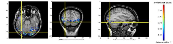
Autism (ASD) is a complex neurodevelopmental disorder that affects social interaction and communication, often impairing individuals for a lifetime. In our study, we used magnetoencephalography (MEG), a non-invasive brain imaging technique, to identify possible biomarkers for ASD. We hypothesized that there would be significant differences in brain connectivity patterns between the ASD group and the controls. We recorded the brain activity of individuals looking at a stationary colorful image while in the resting state. The resting state refers to the brain activity of a subject when he or she is not engaged in any particular task. We found the ASD group had a high concentration of coherent brain activity in the frontal lobe, while the control group had a high level of coherence in the occipital lobe. Areas of high coherence indicate that the brain is well connected and communicating with many other areas of the brain. In controls, we expected high coherent activity in the occipital cortex, since they were looking at a colorful picture. In the ASD group, we found that the frontal lobe was unusually active. This area is typically used in higher-level cognition. These regions of abnormally high coherent brain activity indicate possible biomarkers for autism. Additionally, the ASD group had a significantly lower overall level of coherence than controls.
This article has been tagged with: